
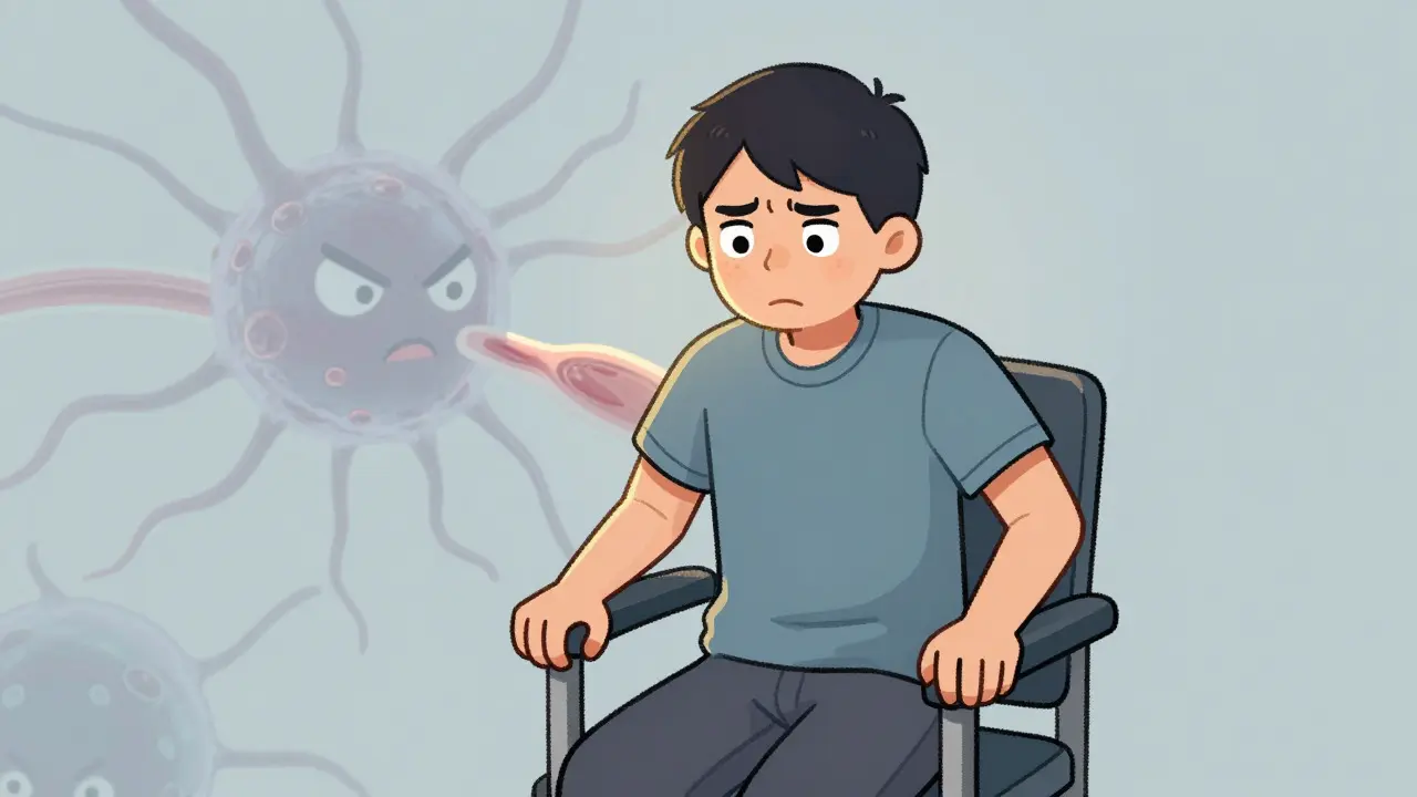
Imagine trying to stand up from a chair or lift your arms to brush your hair, only to find your muscles simply refuse to cooperate. This isn't just fatigue; it’s a signal that something is wrong with the way your body processes movement. For many people, this struggle is the first sign of Dermatomyositis, an autoimmune condition causing muscle weakness and distinctive skin rashes. Or perhaps there are no skin changes, but the progressive weakness in your hips and shoulders remains relentless, pointing toward Polymyositis, a related chronic inflammatory myopathy affecting voluntary skeletal muscles without skin involvement.
These two conditions fall under the umbrella of inflammatory myopathies. They are rare, serious, and often misunderstood. The good news? With early diagnosis and modern therapy, most people can regain strength and live full lives. But getting there requires knowing what to look for, who to see, and how to navigate the complex world of immunosuppressive treatments.
Understanding the Difference: Skin vs. Muscle Only
The core issue in both dermatomyositis and polymyositis is inflammation. Your immune system, which usually fights off viruses and bacteria, mistakenly attacks your own muscle tissue. This leads to damage, weakness, and pain. However, the presentation differs significantly between the two.
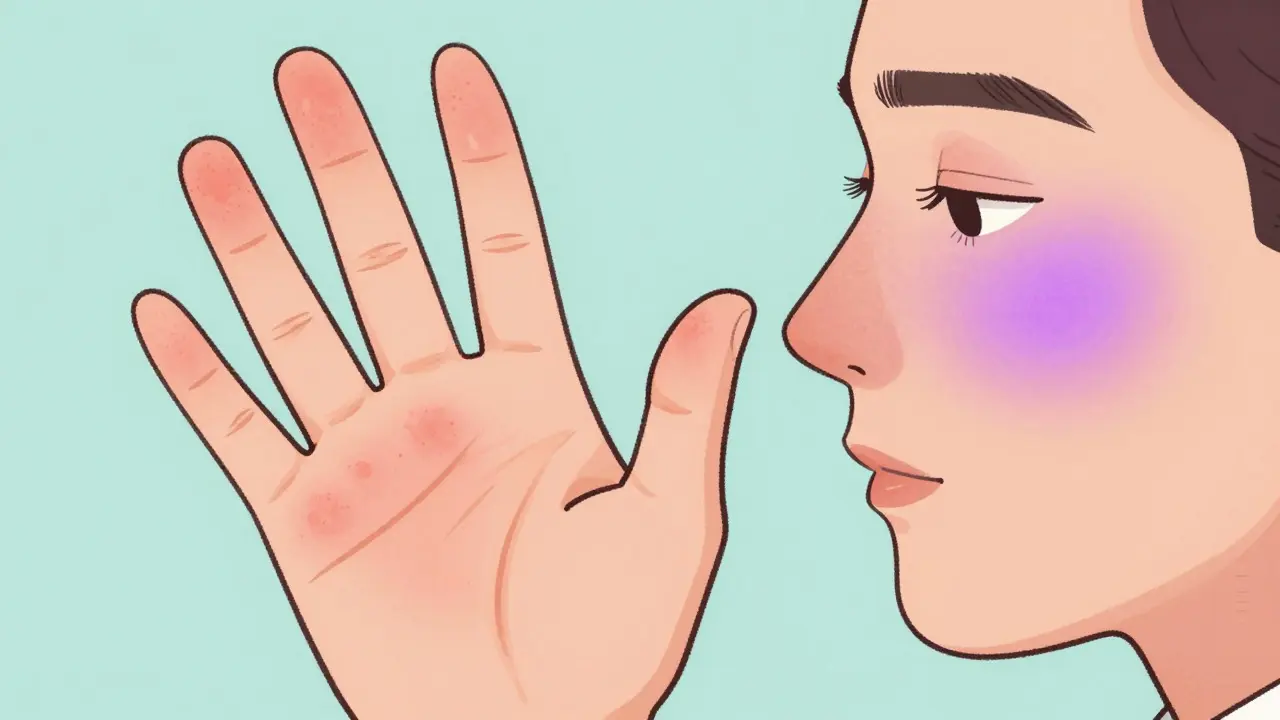
Dermatomyositis is defined by its dual impact. You will experience muscle weakness, yes, but you will also have visible skin changes. A hallmark sign is the heliotrope rash-a purplish discoloration on the upper eyelids, often accompanied by swelling. Other common skin signs include Gottron’s papules, which are red or purple bumps over the knuckles, elbows, and knees. If you notice these specific rashes along with muscle trouble, dermatomyositis is the likely suspect.
Polymyositis, on the other hand, stays hidden beneath the surface. It affects the same proximal muscles-those closest to your trunk, like your thighs, hips, shoulders, and neck-but spares the skin. Because there are no visible clues, polymyositis is harder to spot initially. Both conditions affect women more than men, typically appearing in adulthood between ages 30 and 60, though dermatomyositis can also strike children.
Recognizing the Early Warning Signs
Catching these diseases early is crucial because untreated inflammation causes permanent muscle damage. Here is what you need to watch for:
- Proximal Muscle Weakness: This is the primary symptom. It’s not just feeling tired; it’s mechanical failure. Do you struggle to comb your hair, reach overhead shelves, or rise from a low chair without using your hands? Do stairs feel impossible?
- Dysphagia (Swallowing Difficulty): About 15-30% of patients experience trouble swallowing. This happens because the muscles in the throat and esophagus are also affected. Choking on liquids or food sticking in the chest is a major red flag.
- Fatigue: Over 68% of patients report severe, unexplained fatigue that limits daily activities. This isn’t the kind of tiredness sleep fixes.
- Joint Pain: Arthralgia occurs in many cases, mimicking arthritis.
- Skin Manifestations (Dermatomyositis only): Look for the heliotrope rash on eyelids, photosensitivity (rashes worsening in sunlight), and mechanic’s hands (cracked, rough skin on fingers).
If you have symmetric weakness in your shoulders and hips, especially if it has been progressing over weeks or months, do not wait. See a specialist.
The Diagnostic Journey: Why It Takes Time
Getting a correct diagnosis is rarely straightforward. Studies show that about 30% of patients are misdiagnosed initially, often being told they have fibromyalgia, lupus, or thyroid issues. The average time to diagnosis can take years, involving multiple specialists. To get it right, doctors rely on a combination of tests.
| Test | What It Checks | Typical Findings in Myositis |
|---|---|---|
| Blood Tests (CK Levels) | Muscle enzyme leakage | Creatine phosphokinase (CPK) levels often 5-10 times higher than normal (normal range 10-120 U/L) |
| Electromyography (EMG) | Electrical activity in muscles | Short-duration, low-amplitude signals indicating muscle irritation and damage |
| MRI | Inflammation location | Shows areas of edema and inflammation in thigh and shoulder muscles |
| Muscle Biopsy | Tissue structure | Gold standard: Shows T-cell infiltration in polymyositis; perifascicular atrophy in dermatomyositis |
| Autoantibody Panel | Immune system markers | Presence of myositis-specific antibodies (e.g., Anti-Jo-1, Anti-Mi-2) |
A muscle biopsy is often the definitive test. In polymyositis, pathologists see immune cells attacking muscle fibers directly. In dermatomyositis, the attack targets blood vessels surrounding the muscle, leading to distinct patterns of wasting called perifascicular atrophy. Additionally, because about 20% of adult dermatomyositis cases are linked to underlying cancer (particularly ovarian, lung, or gastrointestinal), a thorough malignancy screening is standard protocol at diagnosis.
Treatment Strategies: Calming the Immune System
There is no cure for dermatomyositis or polymyositis, but the goal of therapy is remission-stopping the inflammation so muscles can heal. The approach is aggressive and immediate.
First-Line Therapy: Corticosteroids
Prednisone is the cornerstone of initial treatment. Doctors typically start with high doses (around 1 mg per kilogram of body weight per day) to quickly suppress inflammation. Most patients see improvement within 4 to 8 weeks. However, long-term steroid use comes with significant risks, including osteoporosis, diabetes, weight gain, and cataracts. To mitigate this, doctors aim to taper the dose as soon as possible, often adding other medications to help reduce the steroid load.
Second-Line Agents: Steroid-Sparing Drugs
To allow for lower steroid doses and maintain remission, rheumatologists add immunosuppressants. Common choices include:
- Methotrexate: Often effective for skin symptoms in dermatomyositis and muscle strength in both conditions.
- Azathioprine: Another common option for maintaining long-term control.
- Mycophenolate Mofetil: Particularly useful for patients with interstitial lung disease associated with myositis.
Advanced Therapies
For refractory cases where standard drugs fail, options include Intravenous Immunoglobulin (IVIG), which provides healthy antibodies to modulate the immune response. Rituximab, a B-cell depleting agent, has shown success in difficult cases, particularly for dermatomyositis. Recent trials also highlight JAK inhibitors (like tofacitinib) showing promise in improving skin scores and muscle strength in resistant dermatomyositis.
Lifestyle Management and Rehabilitation
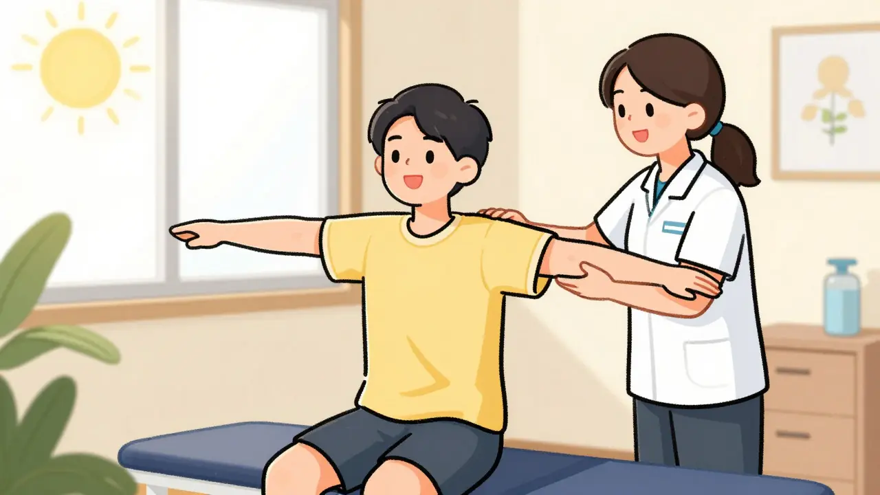
Medication stops the attack, but rehabilitation rebuilds the muscle. Physical therapy is not optional; it’s essential. The key is timing and intensity. Too much exercise during active inflammation can worsen damage, but too little leads to atrophy.
Therapists design low-resistance programs focusing on endurance and gentle strengthening. As inflammation subsides (monitored by falling CK levels), the intensity increases. Many patients report that structured PT improved their functional capacity by over 35% within six months. Speech therapy may be needed if swallowing difficulties persist.
Dietary adjustments also play a role. Since steroids increase appetite and cause fluid retention, a balanced diet low in sodium and high in calcium and vitamin D helps manage weight and protect bone density. Sun protection is critical for dermatomyositis patients, as UV exposure triggers rashes.
Living with Myositis: Prognosis and Outlook
The outlook for these conditions has improved dramatically. Decades ago, survival rates were poor. Today, with modern immunosuppression, 10-year survival exceeds 80% for dermatomyositis and 85% for polymyositis. About 80% of patients achieve remission or low disease activity if treated promptly within the first six months of symptoms.
However, life with myositis requires vigilance. Regular monitoring of CK levels, liver function, and lung health is necessary. Patients must balance medication side effects with disease control. Support groups and patient communities provide invaluable emotional support and practical tips for navigating insurance hurdles and specialist shortages.
Is dermatomyositis or polymyositis contagious?
No. Neither dermatomyositis nor polymyositis is contagious. They are autoimmune disorders, meaning your immune system mistakenly attacks your own body. You cannot catch them from another person through contact, air, or fluids.
Can these conditions go into remission?
Yes. With appropriate treatment, approximately 80% of patients achieve remission or low disease activity. Remission means symptoms are minimal or absent, and muscle strength stabilizes. However, some patients may experience flares, requiring adjustment of medication.
Why is cancer screening important for dermatomyositis?
About 20% of adults diagnosed with dermatomyositis have an underlying malignancy, such as ovarian, lung, breast, or colorectal cancer. The cancer can sometimes trigger the autoimmune response. Therefore, comprehensive cancer screening is recommended at the time of diagnosis and monitored closely for the first few years.
What is the difference between polymyositis and inclusion body myositis?
Inclusion body myositis (IBM) is different from polymyositis. IBM typically affects older adults (over 50), causes asymmetric weakness (often in finger flexors and quadriceps), and does not respond well to immunosuppressive therapies. Polymyositis causes symmetric proximal weakness and responds to steroids and immunosuppressants.
How long does it take for treatment to work?
Most patients begin to see improvement in muscle strength within 4 to 8 weeks of starting corticosteroid therapy. Full recovery takes longer, often several months to a year. Blood tests (CK levels) should drop significantly within the first month if the treatment is effective.




Terrence spry
I'm a pharmaceutical scientist specializing in clinical pharmacology and drug safety. I publish concise, evidence-based articles that unpack disease mechanisms and compare medications with viable alternatives to help readers have informed conversations with their clinicians. In my day job, I lead cross-functional teams advancing small-molecule therapies from IND through late-stage trials.
view all posts