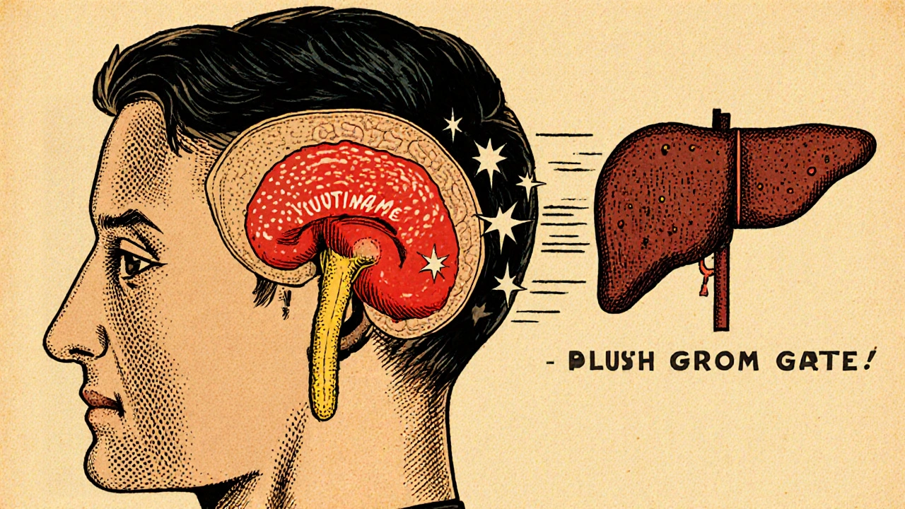
When it comes to acromegaly diagnosis, getting the basics right saves years of uncertainty. Acromegaly diagnosis, the systematic approach doctors use to confirm excess growth hormone production that causes abnormal tissue growth. Also known as acromegaly testing, it blends symptom checks, lab work, and imaging to pinpoint the problem.
The first clue usually starts with physical signs—enlarged hands, facial bone changes, or persistent joint pain. Those signs point to Growth hormone excess, a condition where the pituitary gland releases too much GH, driving tissue overgrowth. To prove that excess, doctors rely on IGF-1 testing, a blood test that measures insulin‑like growth factor‑1, which stays elevated in chronic GH over‑production. The test is a cornerstone because IGF‑1 levels remain high even when GH spikes are brief. If the lab work suggests a problem, the next step is to locate the source. Most cases trace back to a Pituitary adenoma, a benign tumor in the pituitary gland that secretes excess growth hormone. High‑resolution MRI imaging, magnetic resonance scans that give detailed pictures of the pituitary region confirms the tumor’s size and exact position. Together, these steps create a clear chain: symptoms lead to biochemical testing, which directs imaging, which finally identifies the tumor. The relationship between these entities forms the diagnostic backbone. In semantic terms, "Acromegaly diagnosis encompasses biochemical testing," and "Biochemical testing requires IGF‑1 measurement." Likewise, "Pituitary adenoma influences growth hormone excess," while "MRI imaging confirms pituitary adenoma." Understanding these links helps clinicians move swiftly from suspicion to confirmation. Beyond the core steps, a multidisciplinary team often joins the effort. Endocrinologists interpret hormone levels, radiologists read the scans, and surgeons plan potential removal. Early detection matters because untreated acromegaly raises the risk of cardiovascular disease, diabetes, and joint degeneration. By catching the condition through the outlined workflow, patients can access treatments—surgery, medication, or radiation—before complications set in. Below, you’ll find a hand‑picked collection of articles that dive deeper into each part of this process. From detailed explanations of IGF‑1 thresholds to the latest MRI techniques and surgical options, the posts give you practical, up‑to‑date information to guide every stage of acromegaly diagnosis.

A clear, easy-to-follow guide on acromegaly covering symptoms, diagnosis steps, treatment options, and everyday management tips.
read more